Primate Neuropathology
Because of their close resemblance to humans, non-human primates are used as animal models in basic neuroscientific research and for the development of neurological therapies. For this purpose, fundamental knowledge both, of normal neurophysiological processes and their morphologic correlates as well as spontaneous or experimentally induced lesions in the non-human primate brain is indispensable. In the context of our routine diagnostic procedures, spontaneous or possibly induced neuropathological changes are systematically processed and comprehensively characterized. The spectrum ranges from SIV-associated neuropathies (e.g. giant cell encephalitis, cytomegalovirus neuritis) via various infections (viral, bacterial, parasitic, fungal) and other inflammatory-degenerative changes (e.g. leukoencephalopathy, Guillain-Barré syndrome) towards neoplastic processes (e.g. meningioma, cerebral lymphoma). Further, a prospective study within a multidisciplinary senescence project will focus on extensive neurohistological investigations according to age-related changes in the marmoset brain (Callithrix jacchus).
Relevant literature:
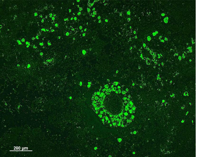
Guillain-Barré syndrome in a rhesus macaque
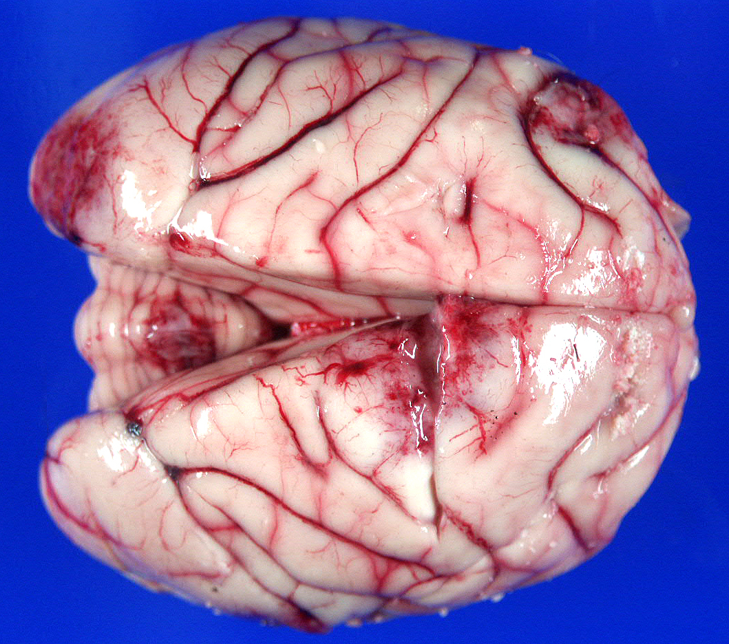
Spontaneous meningioma in a pig-tailed macaque