Magnetic Resonance Imaging

Magnetic resonance imaging and magnetic resonance spectroscopy
The magnetic resonance imaging (MRI) is a modern imaging technique that had become an indispensable part of medical diagnostics and biomedical research. Without any damage to or injuring of tissue, an MRI provides us with high-resolution, high-contrast images of the inside of the intact living organism. In contrast to the X-ray technique, this method is applied without the use of ionizing radiation and is therefore completely harmless for the body.
Besides high-resolution anatomical images, MRIs also provide us with important information concerning the function and metabolism of individual organs. With an MRI we can therefore construct a representation of the blood supply or indirectly examine the changes in the local oxygen supply and activities of the individual brain regions and their interaction. By using the localized magnetic resonance spectroscopy and without extracting any blood or tissue samples, important metabolic products such as sugar and lactic acid and important neurotransmitters of the brain such as glutamate and gamma-aminobutyric acid (GABA) can be determined. All of these studies can be carried out in an almost identical manner in healthy or sick humans and healthy or sick animal, which allows cross-species comparisons.

Magnetic resonance imaging at the DPZ
Since April 2015, the DPZ has an imaging center, which accommodates two magnetic resonance scanners, a cardiac catheter laboratory, offices and animal keeping facilities. The equipment is mainly used for research in neuroscience at the DPZ and thus ensure an optimal extension of the range of methods. In cooperation with the University of Göttingen, a department for functional imaging has been established in July 2015. Head of the new department is Susann Boretius, who was Professor of Biomedical Imaging at Kiel University before. The MRI- equipment consists of 3 Tesla scanners for larger non-human primates and humans, as well as a 9.4 Tesla scanners for small primates and rodents.
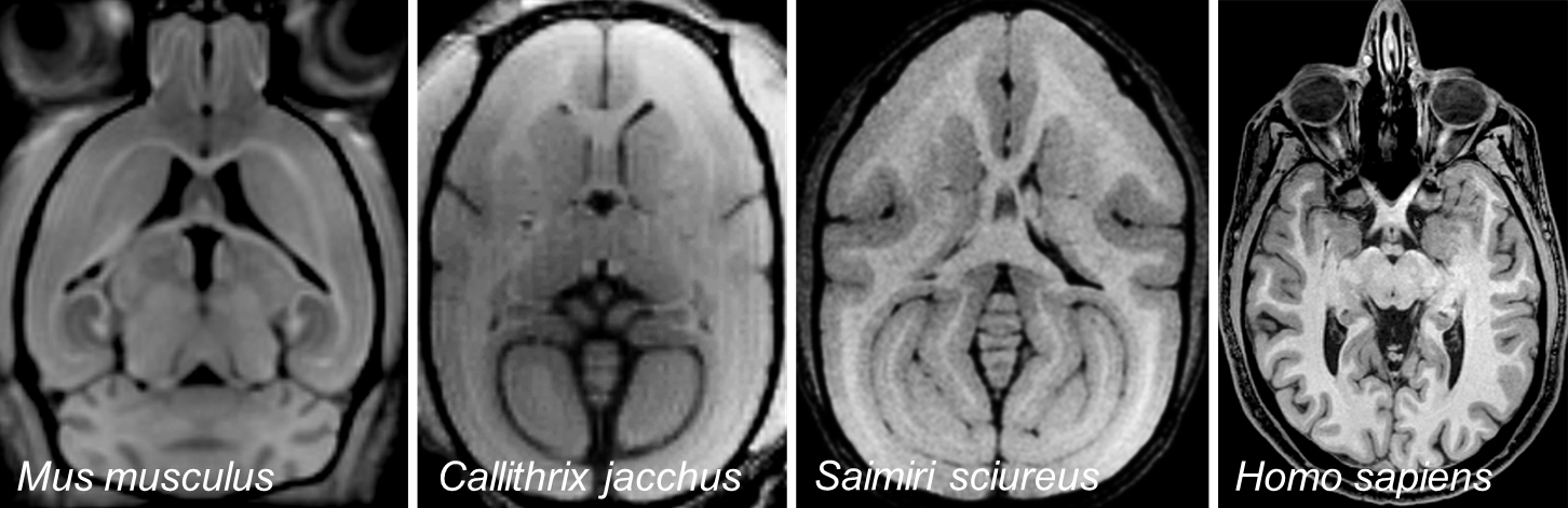
With the implementation of both devices, comparisons between different species from the mouse to humans are possible. The scanners will not only be used by the researchers of the DPZ but also by the scientists of other research institutions of the Göttingen Campus. A cooperation with the German Center for Cardiovascular Disease and the German Center for Neurodegenerative Diseases is planned. In future, the magnetic resonance scanners will also be an attraction during DPZ tours.
Method
The magnetic resonance technology uses the property of certain atomic nuclei to align themselves in strong magnetic fields. Particularly suitable for this are hydrogen nuclei (protons) since they occur in very large numbers and density in all living organisms. The orientation of the nuclei is altered if radio waves are transmitted. They are transferred to a higher energy state. If the radio signals are switched off, the atomic nuclei will return to the low-energy default state. This process is known as relaxation. The speed of the relaxation process is strongly dependent on the respective local chemical environment of the nuclei. This process allows the possibility to distinguish between different tissue structures and is used in MRI to produce high-contrast images of the inside of living organisms. With the assistance of very sensitive antennas, the magnetization generated by the nuclei is measured and converted to images with the help of computationally powerful computers. Through the rapid repetition of this approach, the functions of individual organs such as the beating of the heart or excretion of substances via the kidneys can be examined in time frames.
In addition to the concentration of the hydrogen nuclei and their different relaxation times, further properties of the tissue can be used to analyze not only the anatomical structure but also chemical composition and function. By using this method, we can for example determine the progression of nerve fibers (diffusion-based imaging) from the diffusion of water molecules. The exchange of magnetization between molecules (magnetization transfer techniques) can also be used for the detection of macromolecules (e.g. the myelin of nerve fibers) as well as for the detection of smaller molecules (e.g. amines or amides). Current developments in the field of MR-compatible contrast agents will enable a multitude of new insights into the molecular processes of the intact organism.
Magnetic resonance imaging scanners
The principle component of a magnetic resonance imager is a very strong static magnetic field. In order to achieve high magnetic field strengths, modern magnetic resonance imaging use superconducting electromagnets. By lowering the temperature to -269 degrees Celsius, the current can flow into the conductor windings of the coil with the assistance of liquid helium and without any electrical resistance. With these systems, magnetic fields that are more than 100,000 times stronger than that of the earth are generated. With the magnetic field, the sensitivity of the method increases. The stronger the static magnetic field, the stronger the magnetization through the alignment of the atomic nuclei, which in return improves the spatial and temporal resolution of the measurement. Nowadays, a resolution or detailed accurate images of living organisms are possible and in previously this was only possible through a microscopic examination.
In order to assign the measured signal to a specific area where three-dimensional images of the body are generated, smaller magnetic fields in the three spatial planes (x, y, z) are generated by the so called gradient coils. These must repeatedly be switched on and off and as a result, the typical hammering noise generated during an MRI measurement is created.
Safety
Although the measurement itself is not harmful, ferrous objects in the vicinity of the scanner could due to the strong attraction of the magnet, end up being harmful projectiles. Watches, cell phones, and credit cards are also not permitted in the vicinity of a scanner, since they can get severely damaged. The magnetic field could also cause damage to electronic devices in the body, such as pacemakers or insulin pumps. In addition, metal-containing tattoo inks or cosmetics can sometimes cause significant errors in imaging with a MR scanner. Entering the premises is permitted only after appropriate training or when instructed by skilled personnel.
More on the basic principles of MRI
How can the atoms in our body find orientation to magnetic field

Functional magnetic resonance imaging
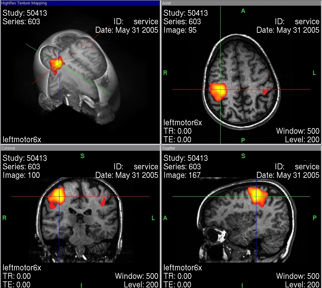
Functional magnetic resonance imaging (fMRI) utilizes the basic method of MRI, to represent physiological functions in certain organs. Examples from the medical practice are the dynamic cardiac MRI or the measurement of the blood sipply of tissue. The most successful applications for functional magnetic resonance imaging lie in the spatial representation of brain activity. The fMRI allows us to visualize changes in blood flow in certain brain areas. These changes are due to increased metabolic activity associated with an increased activity of the nerve cells of the brain. If we for example think of something, look at an object or solve a specific problem, a neural impulse is created at a particular point in the brain. Energy is needed to forward these neural signals through the nerve cells, which in return is reflected in an increase in the metabolism and combines an increased blood supply and oxygen to this part of the brain. More oxygen is needed in the blood for increased metabolic activity. The fMRI uses the so-called BOLD (blood oxygen level dependent)-effect to visualize the difference in oxygen content in the red blood cells. Oxygen-rich blood has a different relaxation time than oxygen-depleted blood and therefore produces a different MRI signal. Elevated oxygen content in the blood is an indication of active nerve cells (in dependence of the strength of the effect red (low activity) until yellow (high activity) in the illustration).
Functional magnetic resonance imaging in brain research
The use of fMRI allows neuroscientists the spatial representation of brain activity in high spatial resolution. For research purposes, images of the brain in an idle state and that of a brain in a stimulated state are compared. The stimulus is often linked to a task that is repeatedly presented to the subjects. Through fMRI recordings, it is possible to visualize the areas of the brain that are important for the processing tasks. The detection of metabolically active regions of the brain enables the assignment and localization of cognitive functions and processes in the brain, for example those areas that are important for certain services such as speech or motor skills. In addition to gaining neurophysiologic information, this knowledge is also of significant importance for the medical practice. Caution must be practiced when brain tumors are removed since it is essential not to damage the area of the brain responsible for language. Neurology and Neuropsychology will also benefit from the use of fMRI. In comparison to healthy people, those who suffer from mental disorders such as depression, anxiety or obsessive-compulsive disorders, show significant differences in brain metabolism. An fMRI can identify the affected regions of the brain.
Magnetic resonance imaging on primates

The MRI measurements at the DPZ are primarily performed with rhesus monkeys. The focus is the research of the cognitive abilities of the brains of primates. In order to be able to obtain high-resolution images and the mapping of certain regions of the brain, the animals are usually anaesthetized (MRI). However, for the visual representation of certain brain activities, it is important that the animals are awake (functional MRI). Rhesus monkeys who participate in neuroscientific experiments in the MRI are trained for several months in advance. During this time, they slowly are acclimatized to the surroundings and the loud noises inside the MRI scanner. To locate the areas of the brain that are important for certain cognitive abilities, the animals must also solve certain tasks during the MRI measurements. The monkeys were presented symbols at regular intervals on a touch screen where they had to detect and press in a specified way. The brain activity is measured while they solve the tasks.
Possibilities and limitations of functional magnetic resonance imaging in brain research

The advantages of the implementation of functional magnetic resonance imaging in brain research are clearly justified in its non-invasiveness. In addition, the body is not burdened by harmful radiation or radioactivity. The high-contrast imaging allows the high resolution and spatial representation of interconnected brain areas that are involved in specific cognitive processes. One of the disadvantages of the fMRI is the fact that only “substitute signals” can be measured in the brain. Instead of specific nerve cell activity, perfusion changes are only obtained in specific brain regions. In addition, by using this method no signals of individual nerve cells can be derived. It merely serves as the capturing of neural mass activity. In order to draw conclusions on the functions of individual neurons, invasive electrophysiological methods such as single-cell recordings are indispensable. Nerve cells communicate via changes in ion fluxes that can be recorded directly as electrical impulses to the cell. The microelectrodes are inserted into the extracellular space and the signals of individual and adjacent cells are measured. Nerve cell areas can be located with accuracy and specific processes, which involve neurons, can be identified. This direct access is not possible with fMRI. However, a major drawback of single-cell recordings is its invasive character. It is much more complex than fMRI and more difficult to replicate. In addition, very little can be said about the dynamics of the neural network. In order to explore the brain in all its complexity and performance, different methods of approach must be combined. Therefore, the functional magnetic resonance imaging is not a complete replacement for invasive testing methods such as single-cell recordings, but this can be a useful addition
Functional Imaging Laboratory
Cognitive Neuroscience Laboratory

Decision and Awareness Group

Research Group Neurobiology

MRI scans
MRI scan of the brain of a rhesus monkey
Frontal MRI scan of the brain of a rhesus monkey (coronal plane). Video: Prof. Dr. Jens Frahm, Biomedizinische NMR Forschungs GmbH, Göttingen
Real time MRI of a beating heart
Real time MRI of a beating heart. Video: Prof. Dr. Jens Frahm, Biomedizinische Forschungs GmbH, Göttingen
fMRI scan of a brain
Horizontal functional MRI scan of a brain (transverse plane). The colored areas indicate increased brain activity, visualized through the BOLD effect. Video: Prof. Dr. Jens Frahm, Biomedizinische NMR Forschung GmbH, Göttingen