Heart patch

Schematic illustration of the application of a heart patch. Figure: Bobbie Smith
Reparing Broken Hearts - Story of a Heart Patch
After 25 years of intensive basic research, the heart patch is scheduled to enter clinical testing for the first time in 2021. A story that could not be told without animal testing and scientific cooperation.
There is good news and bad news. The good: our life expectancy continues to increase with a high quality of life. The bad: Chronic heart disease, and especially heart failure, is already the number one cause of death in the world. With the currently available therapeutic measures, this situation will not change. Rather, the number of patients with heart failure will continue to increase. In 2018, more than 200,000 people died of heart failure in Germany. Wouldn't it be good to be able to repair hearts?
The beginning: rat cardiac muscle cells

This is the challenge that drives the physician Prof. Wolfram-Hubertus Zimmermann - and has been doing so for more than 25 years now. It all began in 1995 with his doctorate at the University Hospital Hamburg-Eppendorf. Still far from actual application in patients, Zimmermann was given the task of developing artificial heart tissue from rat cardiac muscle cells for research into new gene therapy methods. But even then Zimmermann had the idea of direct application in patients with heart failure. And so, a quarter of a century later, the circle has come full circle: if everything goes according to plan, artificial heart tissue from stem cells will be tested for the first time in the first quarter of 2021 as a so-called heart patch in patients with heart failure. But a lot has happened in the meantime, too.
"That's what you can get people excited about. Not for the animal testing, nobody likes to do that, but for the urgently needed development of new therapeutics and diagnostics."

In the mid-1990s, when Zimmermann was a PhD student in Prof. Thomas Eschenhagen's research group, he achieved the first milestone: heart muscle cells from rats formed a three-dimensional tissue that beat spontaneously - just like the healthy heart does. The heart muscle tissue, which is now called "engineered heart muscle" (EHM), was kept alive for about two weeks. This was proof that heart muscle could be produced in the laboratory. The translation of this process into a clinically applicable concept has stayed with Zimmermann ever since. Stem cells as a source of heart muscle cells were needed.
Path blocked by law for the time being
At the end of the 1990s, human embryonic stem cells were developed for the first time and their suitability for heart muscle tissue production was demonstrated by Zimmermann and cooperation partners in Haifa (Israel). Nevertheless, the way for clinical application in Germany remained blocked by law. Socially and politically, against the background of the ethical dilemma associated with embryonic stem cells, the approach via adult stem cells was favored. The introduction of so-called induced pluripotent stem cells from humans, bypassing embryonic stem cells, in 2007 cut the Gordian knot for Zimmermann's research in Germany as well. In 2012, the discoverer of this stem cell technology, Shinya Yamanaka, was awarded the Nobel Prize in Medicine and Physiology. At the same time, this opened the door for the heart patch approach developed by Zimmermann and colleagues to be implemented in the clinic.
Development of the heart patch at a glance
1995: First production of artificial heart tissue from rat cardiac muscle cells.
2000-2006: First evidence of therapeutic application in the rat
2002: First application of human embryonic stem cells for the production of artificial heart tissue
2008-2013: Confirmation of the therapeutic principle in a mouse model
2013: Establishment of a process for the production of clinically applicable human heart muscle preparations
2014: first consultations with the higher federal authority (Paul-Ehrlich-Institut) regarding the path to clinical application
Since 2014: extensive safety pharmacological testing
2021: Start of first clinical trial in patients with myocardial insufficiency (BioVAT-HF; NCT04396899)
Three ingredients are already enough
Cardiac muscle cells and fibroblasts from induced pluripotent stem cells are taken up in collagen and filled into molds of almost any size to produce heart patches with a desired shape and function. To help the heart patches mature, they are mechanically stimulated and then ready for implantation after a few weeks. In 2006, Zimmermann, then freshly appointed junior professor for tissue engineering at the University Medical Center Hamburg-Eppendorf, was able to report for the first time on the successful therapeutic use of the heart plaster in rats. For this, rat heart patches were placed on the damaged heart in the rat model and then sutured.
"You can imagine this a bit like a balloon that you squeeze and it puffs out at a weak spot."
In fact, both a mechanical and an electrical connection was established with the recipient heart, and the sewn-on heart patch was supplied by new blood vessels. In doing so, it fulfills two tasks, Zimmermann explains: "On the one hand, the patch helps the heart to pump sufficient blood through the circulation again by supporting the heart as it beats. But on the other hand, it also strengthens the heart wall on the outside, i.e., thickens it, thus reducing the mechanical stress on the heart wall." Indeed, when the heart wall is weakened from a heart attack, it can become bulging, and blood is no longer forced into the circulatory system but into a "blind sac" called an aneurysm. "You can imagine this a bit like a balloon that you squeeze and it bulges out at a weak spot," the researcher adds.
Animal testing for the urgently needed development of new therapeutics and diagnostics
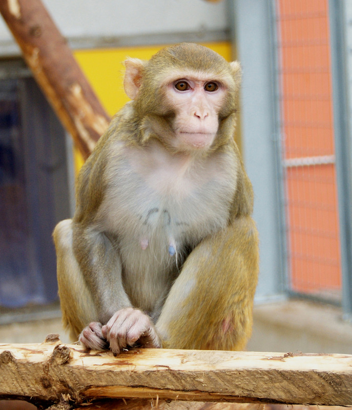
Behr actually prefers to work with common marmosets. However, he was quickly convinced by research partners that rhesus monkeys are better suited for experiments with heart patches. Their hearts are larger and their immune systems more similar to those of humans. In a first series of experiments, healthy monkeys were tested to see how they tolerate the surgery and the application of the heart patch. This was done in cooperation with the clinics for heart surgery at the University Medical Center Göttingen under the direction of Prof. Ingo Kutschka and the University of Lübeck under the direction of Prof. Stephan Ensminger. The animals were then continuously observed and examined with non-invasive imaging techniques. After six months, they were euthanized so that both the body and the hearts could be examined more closely. "Since we were working with stem cells in the graft, we had to rule out the possibility that tumors had formed in the body," Behr said. "Despite a careful search by two pathologists, we couldn't find anything like that," he adds. Also of crucial importance for patients with heart failure is that implantation of heart patches does not result in any negative impairment of cardiac function, for example, due to cardiac arrhythmias.
"The observation that there are no cardiac arrhythmias after heart patch implantation was seminal to the clinical trial that is now underway."
A side effect that cannot be studied in cell culture or in alternative small animal models (e.g. mice). "The observation that there are no cardiac arrhythmias after heart patch implantation was groundbreaking for the clinical trial that is now pending," Zimmermann confirms. This paved the way for the next step of testing the heart patches in monkeys with heart failure. For this purpose, a heart attack was induced in the test animals under general anesthesia in the cardiac catheter laboratory of the German Primate Center, which was established through the German Center for Cardiovascular Research (DZHK). A balloon catheter measuring just a few millimeters is used to occlude a coronary vessel and then reopened after three hours. "As in humans, this results in the death of heart muscle tissue with a reduced pumping function of the heart " explain the Göttingen scientists. After a longer observation phase under intensive veterinary care and clearance by the competent animal welfare authority in each individual case, a heart patch was then implanted to compensate for the loss of heart muscle cells.

After these trials also provided promising indications of stabilization of the heart wall by the implant, the heart patches are now to be tested on humans for the first time. The first step is to identify patients with severe heart failure, who often have no treatment options other than organ transplantation. With only about 300 patients who can be treated with a heart transplant in Germany each year, and against the background of the large number of patients with heart failure, Zimmermann initially assumes that there are about 10,000 potential candidates for the heart patch. If successful in this patient population, however, an expansion of the indication seems possible.
Heart patch as test model reduces number of animal tests
Would the development of the heart patch have been possible without animal testing? "Absolutely not," Zimmermann states. "The crucial thing before the clinical application of the heart patches is, in particular, the certainty that the patient will not be harmed. The tests required for this purpose, which are also prescribed by law, must be tested with the necessary care in a model similar to a human being. A simulation on the drawing board or experiments in the culture dish are helpful and have also been carried out extensively by us, but are not sufficient to be able to make conclusive predictions about the risk potential in humans." On the basis of the data now available, Zimmermann and his colleagues were now in a position to submit a clinical trial application to the higher federal authority, the Paul Ehrlich Institute, to carry out the world's first trial of the heart patch approach in patients. But the heart tissue manufacturing process now developed and continuously optimized by Zimmermann and colleagues also has additional benefits for applications in the laboratory: It helps reduce animal testing in other areas. "Heart preparations along the lines of our heart patch are now widely used as a replacement procedure for animal experiments in drug testing," he says.
"We've always been open about our approaches, and here we can clearly demonstrate how animal testing can contribute to the development of novel drugs."
This makes it possible to test the effect and side effect of drugs on the human heart already in the culture dish and thus optimize them for clinical application for the patient," the researcher reports. Zimmermann is also currently using human heart muscle preparations in collaboration with Prof. Pöhlmann at the German Primate Center to investigate the effect of COVID-19 infection on the human heart as well as new treatment options, for example to prevent sudden cardiac death in the case of SARS-CoV-2 infection. In the process, he also hopes to encourage colleagues to communicate more openly - and to get society excited about scientific topics. "We've always been open about our approaches, and here we can clearly show how animal testing can contribute to the development of novel drugs. That's what you can get people excited about. Not for animal testing, no one likes to do that, but for the urgently needed development of new therapeutics and diagnostics."
Author: Peter Kohl / Source: Tierversuche verstehen